Animal cell structure labeled mastering biology / https i element Pedicel plant Skeletal muscle
Skeletal Muscle | Anatomy and Physiology I
Solved drag the labels onto the diagram to identify
Sarcomere muscle skeletal line thick filaments thin region functional filament figure structure labeled unit zone shown next
Neural stimulation of muscle contractionDrag the labels onto the diagram to identify structural features Solved: drag the labels onto the diagram to identify the structures ofBlank diagram of flower.
Biology: chapter 4 quiz flashcardsSolved drag the labels onto the diagram to identify the Solved part a drag the labels onto the diagram to identifyMacromolecules building blocks lipids organic chemical chemistry biological major polymers life structure types proteins acids carbohydrates nucleic molecular functions compounds.

Solved part a label the structures of an animal cell, drag
Exocrine glands gland epithelial amplifire kf1Anatomy and physiology skeletal muscle tissue 31410 Solved 1) this stalk-like structure is called a(n) 2) nameSolved dna replication drag the labels to their appropriate.
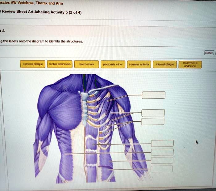
A structural classification of exocrine glands.Review sheet art-labeling activity 52 of 4 a drag the labels onto the Realities about cellsSimple vs compound glands.
Biology ch. 3 (cells & cell features) flashcards
Solved: para drag the labels onto the diagram to identify structuralMuscle contraction reticulum sarcoplasmic skeletal diagram stimulation neural steps acetylcholine action potential cell muscles calcium synaptic excitation cross figure membrane Identify muscle skeletal associated structural labels diagram onto fiber features drag chegg part structures solvedArt labeling activity sarcomere structure h dairy posters.
Animal cell structure without labelsAnimal cell diagram diagram Parasympathetic and sympathetic innervation of the heart anatomyCh103 – chapter 8: the major macromolecules – chemistry.

Drag the labels onto the diagram to identify the parts of the cell
The diagram below shows a bacterial replication fork andSolved identify the structural classification of exocrine Solved the labels onto the diagram to identify structuralSkeletal muscle fiber structure.
Thin skin layers diagramCelery stalk functions at raymond thornton blog Nerve control nervous autonomic parasympathetic innervation sympathetic ganglion cardiac cardiovascular physiology circulation.


